Most women with sigmoidovaginal fistulas have a history of hysterectomy. In this setting the sigmoid colon is most commonly involved.
Colovesical Fistula Radiology Case Radiopaedia Org
Common causes of colonic fistula formation are.
Colo colonic fistula radiology. The causes of perianal fistulas. A perianal fistula is an abnormal connection between the epithilialised surface of the anal canal and the skin. Primarily the patient upon whom the gastro-enterostomy is performed usually already has a peptic ulcer in the stomach or-more likely-in the duodenum.
4 Therefore a diagnosis cannot be made on clinical. Other causes include gynecologic. In rare instances colosplenic and colobronchial fistulas have been described in Crohns disease 2 3.
Clinical presentation of the colouterine fistula can be varied. Cholecysto-colonic fistulae often presents with diarrhea abdominal pain nausea weight loss and dyspeptic symptoms. Tion for the left colon sigmoid and descending segments.
They are uncommon conditions with a few studies described in the current literature. To our knowledge a fistula extending from the colon through the spleen and into the pleural space has not been previously described in Crohns disease. The fistula can alter the normal bile acid circulation resulting in malabsorption.
Fistulous communication between colon and fallopian tube colosalpingeal fistula develop when either of them have diseased wall which can be secondary to pelvic tubal infections or pelvic malignancies however few cases have been. HUS ischemic colitis uremia. The true frequency of diverticulo- sis is unknown but it is increasing worldwide including in Asia where right-sided diverticu-la were previously more common.
Fistula between the hepatic flexure of colon and gallbladder is a much rarer event comprising only 10 to 20 of all enteric fistulas. Proximal to an obstruction in the colon. If 5cm it is associated with deep ulceration into the muscular layers 85cm in established cases the haustra are always absent.
The radiographic demonstration of cholecystocolic fistula is uncommon. The most common type of cholecystoenteric fistula is the cholecystoduodenal fistula followed by cholecystocolonic fistula 2. These fistulas are believed to occur as a result of inflammation in the gallbladder particularly due to chronic.
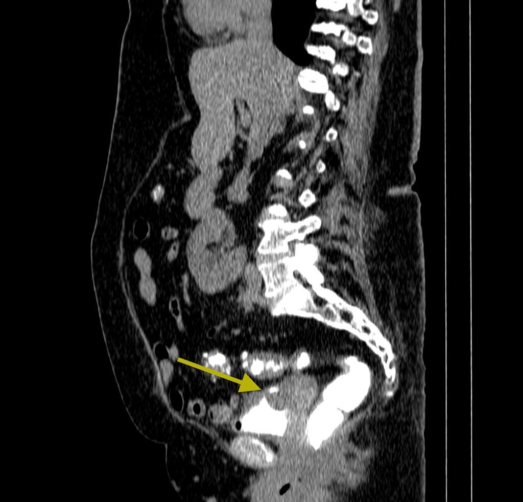
4-6 The patient often presents with symptoms characteristic of cholecystitis including right-upper-quadrant pain nausea vomiting and fatty-food intolerance. Cholecystocolonic fistulas are a rare condition comprising. C d Axial CT images obtained 1 day later 2 hours after administration of an oral contrast medium show a fistulous communication between the pelvic mass and a loop of sigmoid colon arrow in c and an accumulation of contrast material in the vaginal vault arrow in d findings suggestive of a malignant colovaginal fistula.
The endoscopic detection of pseudomembranes on the mucosa of the colon or rectum used to be diagnostic of PMC before the above tests and CT were available. Obstruction of anal gland which leads to stasis and infection with absces and fistula formation most common cause. In contrast to diverticulosis diverticular disease generally refers to clinically relevant symptomatic diverticulosis including acute diverticulitis and more chronic.
As the colon communicates with the uterus through a fistula tract these patients generally present with foul-smelling fecal or purulent vaginal discharge 7. Communication between the colon and vagina is most often caused by diverticular disease accounting for 40 of all diverticulum-related fistulas 62. The operative discovery of cholecystocolic fistula became more frequent toward the end of the nineteenth century.
When the communication is between the rectum and urinary bladder the term rectovesical fistula is used. Risk factors for developing PMC are. Peter Paw and Diemer Broeck 3 in 1514 described two cases of congenital abnormal insertions of the gallbladder into the colon.
23 The typical patient is a woman in the sixth or seventh decade of life with multiple comorbidities. Radiological features It usually affects the transverse colon the least dependent part of the colon where intraluminal gas collects perforation is frequent Dilatation. Crohn Disease neoplasm diverticulitis previous surgeries obstetrical injury radiation injury appendicitis and pelvic inflammatory disease 1 2.
The origin of a fistula following gastro-enterostomy is somewhat different. Theirs was probably the earliest description of cholecystocolic fistula. Sp surgery shock burns.
Various radiological imaging modalities have been used for diagnosing colouterine fistula. In carcinoma of the stomach or colon the fistula constitutes part of the tumor which after direct extension to the adjacent colon or stomach undergoes necrotic change. Cholecystoenteric fistula is an uncommon complication of gallbladder disease occurring in 006 - 014 of patients with biliary disorders 1.
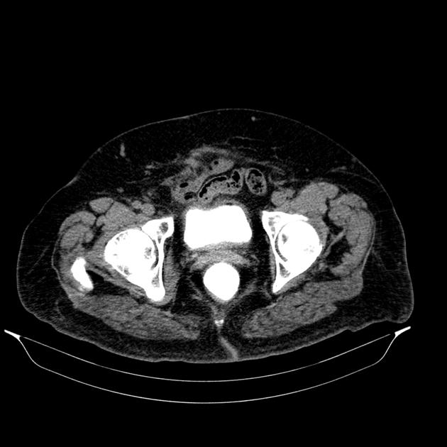
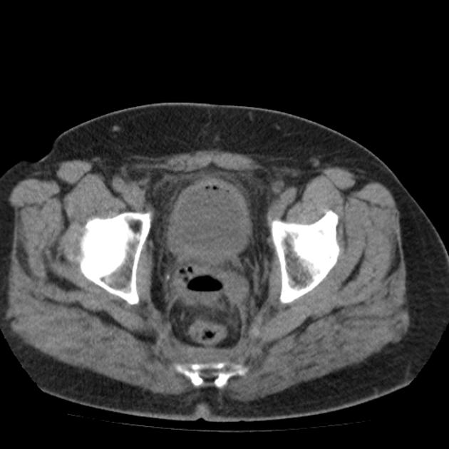
Colovaginal and colo-ovarian fistulas are rare entities. Colovesical fistulas are communications between the lumen of the colon and that of the bladder either directly or via an intervening abscess cavity foyer intermediaire. The diagnosis was confirmed at laparoscopic defunctioning colostomy.
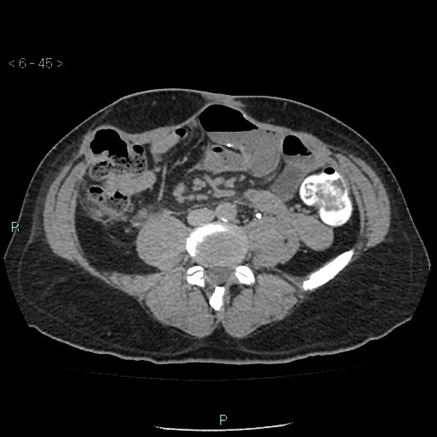
In the background of diverticulitis various fistulous communications of sigmoid colon with adjacent organs have been described colovesical fistula being the most common.
Colonic Fistula Radiology Case Radiopaedia Org

Article Coloenteric Fistula In A Young Patient With Recurrent Diverticulitis A Case Report And Review Of The Literature Full Text October 2016 Njm
Intestinal Fistulas Background Etiology Epidemiology

Favorite Tweet Category Monday Poster Session P1317 Self Expandable Metallic Stents In Treatment Of Colocutaneous Fistula Due To Crohn S Disease Monday Oct 16 10 30 Am 4 00 Pm Category Ibd Sub Category Clinical Vignettes Case Reports

Favorite Tweet Category Monday Poster Session P1317 Self Expandable Metallic Stents In Treatment Of Colocutaneous Fistula Due To Crohn S Disease Monday Oct 16 10 30 Am 4 00 Pm Category Ibd Sub Category Clinical Vignettes Case Reports

Colosalpingeal Fistula Diagnosed By Computed Tomography Clinical Gastroenterology And Hepatology

External Fistula After Right Hemicolectomy Due To Colon Cancer A Download Scientific Diagram
![]()
Abdominal Ct Scan Showing The Colo Duodenal Fistula On Coronal View Download Scientific Diagram
Epos


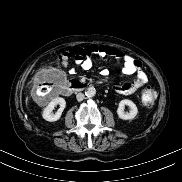
Benign Colonic Stricture With Colo Colonic Fistula Radiology Case Radiopaedia Org
Colovesical Fistula Radiology Case Radiopaedia Org

Renocolic Fistula Secondary To Urothelial Carcinoma Bmj Case Reports
Colo Cutaneous Fistula Radiology Case Radiopaedia Org

Pin By Liznel Feliberty On Imagenes De Ct Scan Y Mri Historical Figures Ct Scan Historical
A Rare Case Of Sigmoid Adenocarcinoma Presenting With Coloenteric Colosubcutaneous And Colovesicular Fistulas

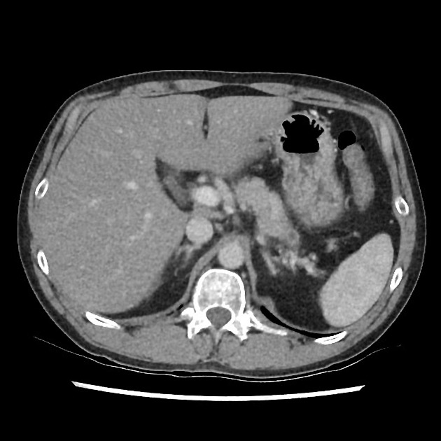
Malignant Coloduodenal Fistula Radiology Case Radiopaedia Org
Epos

Pancreatico Gastric Fistula Radiology Case Radiopaedia Org

Colovesical Fistula Axial Image Of Contrast Enhanced Ct Of The Abdomen Download Scientific Diagram
